The National Library of Medicine has a splendid manuscript collection, including an 18th century Persian text with illustrations. Here is the information from the webpage, with two of the illustrations provided here.
Anonymous Persian Anatomical Illustrations. [Iran or Pakistan, ca. 1680-1750].
Anonymous Persian Anatomical Illustrations.The National Library of Medicine owns approximately 300 Persian and Arabic manuscripts dating from the eleventh to the nineteenth centuries. Most of these manuscripts deal with medieval medicine and science and were written for learned physicians and scientists. Among them are a number of anonymous anatomical treatises or groups of anatomical drawings.
The two featured here consist of a Persian bloodletting figure and a venous figure, probably drawn in the 18th century but based on earlier models (MS P 5 fol. A); and six early-modern anatomical drawings showing some European and Indian influences (MS P 20, item 2).
Six Early Modern Anatomical Illustrations
Six anonymous anatomical drawings occur on folia 554-559 at the end of a volume containing Tibb al-Akbar (Akbar’s Medicine) by Muhammad Akbar, known as Muhammad Arzani (d. 1722/ 1134) in an undated copy probably made in the 18th century. The paper on which these figures are drawn, however, is distinct from that of the main text, though similar in many respects. The illustrations appear to be unrelated to the accompanying text and to draw upon Indian and early-modern sources.
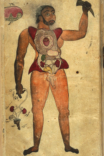
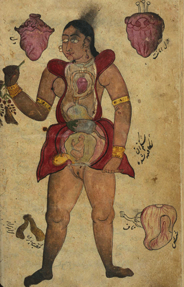
One full-opening of the manuscript, folia 554b-555a, contains two full-figure anatomical illustrations, one of a female and one of a male. The female figure is of a pregnant woman. The woman holds back a flap of abdominal skin to expose the gravid uterus, while in her other hand she appears to hold a plant rather than a part of the body, though that could be interpreted as referring to the female genitalia. Surrounding the figure are portrayals of individual organs: at the top, two hearts; lower right, the lungs; something unidentified in lower left (labeled the opening of the vagina).
The male figure has his abdomen and chest opened to reveal the internal organs. His right hand holds a second set of genitalia, and there is a sketch of the liver and gallbladder in the upper left corner. The artistic conventions employed in the production of these two illustrations clearly indicates Western India as a place of production. The 16th to 18th-century European convention of picturing partially-dissected bodies as if they were alive, often with the obliging cadaver holding up parts of their own body for further inspection, can be seen here transferred to the Indian subcontinent. The anatomy of the exposed organs reflects indigenous Indian concepts as well as some medieval Galenic anatomy.
A second full-opening of the manuscript, folia 556b-557a, contains drawings of individual organs drawn in inks and opaque watercolors. On the righthand page are the liver with gallbladder, the stomach with intestines, the testicles, and a detail of the stomach. On the left are a composite rendering of the tongue, larynx, heart, trachea, stomach, and liver; a composite drawing of the ureters, urethra, kidneys, testicles, and penis; the external female genitalia; and a composite rendering of the internal female genitalia with a gravid uterus. These leaves of individual organs show considerable influence from early-modern European anatomical atlases, while the explicitness of many of the details reflects Indian artistic conventions not found elsewhere in the Islamic world.
A third full-opening of the manuscript, folia 558b-559a, has two drawings in ink and light-gray wash of skeletons, one leaning on a pedestal and the other leaning on a scythe. These two figures are clearly derivative from Vesalian illustrations.
